Analysis and Visualisation Software / TMS Neuronavigator
TMS Neuronavigator
TMS Neuronavigator is a hardware and software system which enables the precise and individual navigation of a TMS coil above a specific anatomical area of the brain, as well as an imaging-guided navigation of the TMS coil to functionally defined brain Regions-of-Interest. The system contains the BrainVoyager QX software package, a special neuronavigation software module and the digitizer hardware. The TMS Neuronavigator system can be used with any TMS coil. The Magstim figure of eight coil and the Medtronic MC-B70 coil are directly supported.


Transcranial Magnetic Stimulation (TMS)
Transcranial Magnetic Stimulation (TMS) is a well-established tool for inducing transient changes in brain activity non-invasively in conscious human volunteers. An increasing number of studies have used the behaviour-modulating capacity of TMS to reveal causal brain-behavior relationships, investigating an ever increasing range of different behavioral, cognitive, and affective functions. Recently, TMS has also been applied as a therapeutic tool for treating a variety of neurological and psychiatric disorders including, for example, depression, neglect, stroke, or schizophrenia. All these studies clearly demonstrate the potential of TMS to serve as a unique research tool for the investigation of a broad variety of issues in cognitive neuroscience. Different experimental TMS protocols can be designed to address questions concerning the location, timing, lateralization, functional relevance or plasticity of the neuronal correlates of information processing. The hypotheses underlying these different experimental TMS protocols can be based on respective results of functional imaging studies, neuropsychology, or animal models, investigating the same paradigms and neuronal pathways from methodologically different perspectives. The increasing use of TMS in Cognitive Neuroscience and Neuropsychiatry requires the precise positioning of the TMS coil above a specific anatomically or functionally defined brain region, individually for every participant or patient. Only individual imaging-guided neuronavigation of the TMS coil is capable of achieving the precision in positioning the TMS coil, necessary for successful and reliable TMS experiments and clinical treatments.
How Real-Time TMS Neuronavigation Works
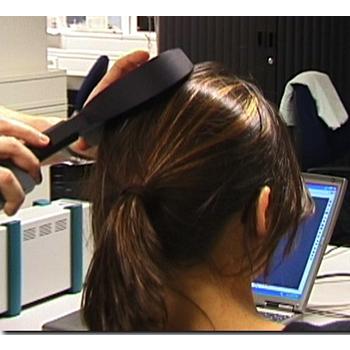
During TMS Neuronavigation, stereotaxic data for the localization of the TMS stimulation site are recorded using an ultrasound-based co-registration system. This system consists of several miniature ultrasound senders which are attached to the participant's head and the TMS coil. These ultrasound senders continuously transmit ultrasonic pulses to a receiving sensor device. The measurement of the relative spatial position of these senders in 3d space is based on the travel time of the transmitted ultrasonic pulses to three microphones built into the measurement device.
In the next step, local spatial coordinate systems are created by linking the relative raw spatial position of the ultrasound senders to a set of fixed additional landmarks on the participant's head. The specification of these fixed landmarks is achieved via a digitizing pen that also hosts two transmitting ultrasound senders in order to measure its relative position in 3d space. The nasion and the two incisurae intertragicae are used as the three anatomical landmarks in order to define the local coordinate system. After this stage, the system provides topographic information of the head ultrasound senders relative to a participant-based coordinate frame. Similarly, the TMS coil also hosts a set of ultrasound senders whose relative spatial positions are linked to fixed landmarks specified on the coil in order to calculate another local coordinate system.
Once the local spatial coordinate system is defined for the participant's head and the TMS coil in real 3d space, these coordinate systems have to be co-registered with the coordinate system of the MR space. For TMS-MRI co-registration, the same digitized landmarks on the participant's head are specified on the head representation (mesh) of the participant in the neuronavigation software. The anatomical landmarks are identified in the MRI scan of the participant's head and co-registered with the coordinates from the digitizer. After the landmarks specified on the real head are co-registered with those on the mesh head, movements of the TMS coil relative to the head of the participant in real space are registered online and visualized in real-time at correct positions relative to the participant's anatomical reconstruction of the brain.
By superimposing the functional data on the anatomical reconstruction of the brain, the TMS coil can be neuronavigated to a specific anatomical and/or functional activation area in every participant. TMS neuronavigation should always be based on data in AC-PC space (rotating the cerebrum into the anterior commissure – posterior commissure plane) in order to avoid any additional transformations that could distort the correspondence between MRI and stereotaxic points. For structural and functional MRI analysis, the full power of BrainVoyager QX is available.