Analysis and Visualisation Software / BESA EEG/MEG Analysis
BESA EEG/MEG Analysis
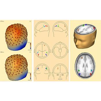
Versatile and user friendly signal processing for EEG and MEG data. The most widely used software in EEG and MEG research, developed on the basis of 20 years experience in human brain mapping. Wide variety of source analysis algorithms. Bidirectional connection with BrainVoyager™ allows source seeding from fMRI clusters with one mouse click


Installation
How do I install BESA on my computer?
Install from your installation CD. A complete BESA installation can also be downloaded from the link Updates/BESA.
How do I install BESA updates on my computer?
Download the self-extractable update file from our website and expand over the existing installation.
Can I run BESA on a Linux computer?
Yes. Because BESA was designed specifically for Windows, this requires an emulation software like VMWare and a legal license of Windows installed on the computer. BESA will then run with LINUX hosting Windows in the virtual environmernt of VMWare. For further instructions, please refer to our manual Running BESA under Linux.
How do I upgrade from version 4.x to version 5.1.x?
Install the new BESA CD version 5.1.x and obtain the electronic hardlock update file from our sales office if you have a BESA hardlock with serial number. Note: Users with BESA99 hardlocks without serial number need to purchase the upgrade with a new hardlock.
How do I install or update the hardlock driver?
BESA requires a hardlock and the correct driver. When installing from CD, the hardlock driver is installed automatically. For updates see ReadMe.txt in BESA\Utilities\Hardlock for more details.
I installed BESA on Windows XP, but I cannot start the program
Please download the latest hardlock driver 'hldrv32.exe' from the Aladdin webpage
What is a floating network license and how does it work?
A floating network consists of one server and several client computers. Within the network, a specific number of users can run BESA simultaneously from any client and/or server computer in your institution. The number of users is determined by the internal coding of the network hardlock which is connected to the server PC. See ReadMe.txt in BESA\Utilities\Hardlock for more details.
Can the BESA server of a floating network be a normal PC?
Yes. The server for a floating network can be any PC within the network. The hardlock service and a monitor program have to be installed on the BESA server computer. The server PC should be running continuously. If the server is rebooted, the hardlock service starts automatically. The environment settings of the client computers in your institution may have to be set specifically to find the server computer. See ReadMe.txt in BESA\Utilities\Hardlock for more details.
I installed the latest BESA update from your website but cannot start the program.
New updates contain only the additional files for the update version. Therefore please make sure not to remove the older version before running the update and select the correct drive/folder for expanding the update into.
In my BESA directory of version 4.2, I find different executables (BESA.exe, BESA2000-EEG.exe and BESA2000-MEG+EEG.exe). What is the difference between them and which one am I supposed to use?
If your hardlock is labeled BESA99-EEG, please use BESA2000-EEG.exe. If your hardlock is labeled BESA99-MEG, please use or BESA2000-MEG+EEG.exe. BESA.exe is used with the new BESA 2000 hardlocks. The numbered BESA-2000-xxxx hardlocks can be upgraded electronically
Data Analysis
Which EEG and MEG file formats are supported by BESA?
Click here to download a list of supported file formats.
What is the best strategy to design a multiple source model?
The best strategy is usually a sequential spatio-temporal approach. However, this may depend on the data to be analyzed. For strategies and examples, please read the key papers on multiple source analysis in Publications / Methods.
How can I handle artifacts in BESA?
It is important to get good templates for the topographies of the artifact using pattern search and averaging. Then the topographies can be used as spatial components during source analysis to separate brain from artifactual activities (preferred method). For this load the data segments and the averaged artifacts into the source analysis module and use the PCA on the artifacts to add spatial components to the solution. The second method is based on subspace projection which regressed the artifact topographies out and distorts the topographies during source analysis. Use the artifact correction of the on-going EEG, average and save the corrected averages. When reopening a corrected average file, artifact correction coefficients will be read and automatically used (only in BESA 5.0) for subspace proejction of the data prior to source analysis. In the source analysis module, the forward vectors will be subspace projection and (similarly) distorted. The advantage of the first, preferred method ist that the source waveforms of the artifact and brain signals can be compared to see the epochs of critical interference. Maps are not distorted. The artifact correction of the on-going EEG (second method) contrast the artifact against the background EEG rhythms but not against the much smaller evoked response components to be averaged.
How do I export dipole moments of my fitted sources?
The source waveforms contain the dipole moments of the sources calculated for each time point. Source waveforms can be written to an ASCII file: Use the 'File | Save Source Waveforms As...' menu entry.
Other Questions
What kind of electrode configurations can be recommended?
For a detailed description, please see the document Recommended electrode configurations. There are three important aspects for source analysis and source montages:
Electrodes should have equal spacing. A row of inferior temporal electrodes on both sides is important to cover the lower head sufficiently, e.g. F11 (F9), A1, P11 (P9). The lower sites (F11, P11) at a 20% distance are preferable to sites at a 10% distance (F9, P9) from the standard row F7, T7, P7 (same applies to other side).
Electrodes should be symmetric between both hemispheres with a midline reference or an implicit reference (e.g. F3+F4). A recommended standard with 33 electrodes adds a midline reference Pz, and F11, A1, P11, FC1, FC5, CP1, CP5 and their homologues over the right hemisphere to the 19 electrodes of the 10-20 system (Fpz, Oz not included). For more details see BESA help chapter on Electrodes. When using caps with more electrodes (64 or 128) inferior electrode and equal spacing are similarly important.
How can I verify that electrode locations were imported correctly?
The electrode locations can be inspected in a 3D view using the menu command 'File|Head surface points and sensors|View'. The fiducials (pre-auricular points and nasion) are plotted as purple cubes, the electrodes are plotted as red disks. By clicking on a disk, the electrode label of a particular location, and its coordinates, can be checked. The calculated head radius is also given (default value is 85 mm if no electrodes were digitized).
How do I import digitized electrode locations and head surface points?
Digitized electrode locations can be provided in a surface point file (extension *.sfp) in ASCII format. It is recommended that the first three digitized coordinates are the fiducials (fiduciary points), labeled 'FidT9', 'FidT10', FidNz', followed by electrodes, and possibly additional skin points. A detailed description of the file and how to import it is given in the BESA help files (chapter: Working with Electrodes and Surface Locations).
How do I change or exclude erroneous head surface point locations?
After importing head surface points, they can be inspected using the menu command 'File|Head surface points and sensors|View'. If some points were not digitized correctly, it should be apparent from the 3D view. To exclude these points, the head surface points can be written to an ASCII file by using the command 'File|Head surface points and sensors|Save all files in head coordinates'. The digitized head surface points are written to a file entitled 'xxx.sfp'. They can be edited using a text editor program. Then the data file can be re-opened in BESA, and the edited head surface point file is read automatically. Important note: When using MEG and EEG simultaneously, it is important that the 'xxx.pos' file is also written and read in the same coordinate system as the surface points!
How do I import digitized electrode locations in combined EEG / MEG measurements with the Neuromag system?
The Neuromag system does not store the electrode names with the coordinates. Thus, it is extremely important that you keep track of the digitization protocol, and provide a file which provides the names of fiducials, electrodes, and additional head surface points in the order of digitization (see BESA help: Working with Electrodes and Surface Locations).
How do I import Top Viewer waveforms into a graphics software for further processing?
Top Viewer waveforms can be exported into encapsulated post script files using the command 'File | Print to File' in the Top Viewer window. Printed files can then be imported into a graphics software (e.g. Corel Draw, Adobe Photoshop). In vector-oriented graphics programs, the scalability of the eps format remains available if you select 'Post Script Interpreted' when importing.